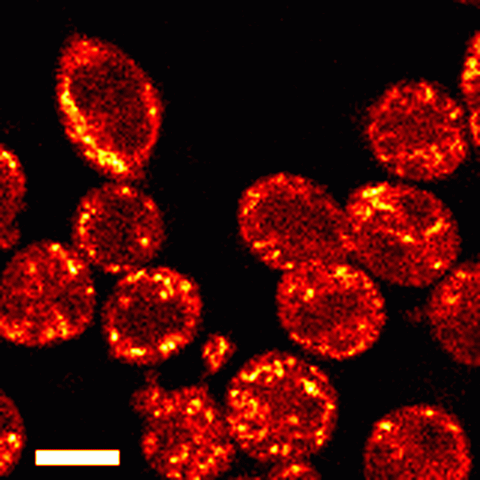
The fluorescence of intrinsic cellular fluorophores can be used to reveal the cellular metabolic state. This pictures shows NADH fluorescence from rat basophillic leukemia cells excited using ultrashort pulses of light at 740 nm. The bright, punctate regions are individual mitochondria (scale bar = 10 microns). By calibrating the fluorescence from normal cells against the fluorescence from cells that have been treated either to inhibit cellular respiration or to uncouple oxidative phosphorylation from electron transport, a quantitative measure of the cellular energy state can be formed.
Recent experiments in the mouse cochlea have revealed that in addition to NADH, flavoprotein fluorescence can also be detected and quantified. Our current research efforts are aimed at using the Redox state of NADH and flavoprotein to determine spatial and temporal energy gradients in inner and outer hair cells of the organ of Corti.


